


The 2020 AHA guidelines for ACLS recommend using quantitative waveform capnography in patients during CPR. To better understand how to use waveform capnography for more effective resuscitation efforts, we will examine the subject in detail and its application to ACLS scenarios.
What is Capnography?
Capnography is a non-invasive method for measuring the partial pressure of CO2 from the airway during inspiration and expiration. A sensor is used that detects expired CO2 levels, which is attached to a bag mask device or endotracheal tube (ET tube).
Capnography readings provide information on ventilation, perfusion, and metabolism – which are all important for successful airway management.
There are two types of CO2 measurement in use: capnometry and capnography.
- A capnometer is a device that provides a numeric CO2 reading of exhaled gas. This measurement is called end-tidal CO2. It is helpful for measuring the success of treatments, effective ventilations, and for monitoring respiratory conditions like COPD. Normal adult patient CO2 values range between 35-45 mmHg, or at about 5% CO2.
- A quantitative waveform capnography reading provides more information than a capnometry reading alone, because it shows the entire inspiration-expiration cycle of each respiration as well as the end-tidal CO2 reading. A capnography waveform represents air movement in the lungs — very much in the same way an ECG represents electrical conduction in the heart.
Per AHA recommendations for ACLS, the focus of this article will be on waveform capnography rather than capnometry, although this method does have value in other care scenarios.
History of Capnography
Capnography began to be used in the clinical setting during World War II, first in the Netherlands. It was noted that adverse events can be avoided by using capnography as patients were awakening from anesthesia. Patients who seemed awake could still be under the effects of muscle relaxants and therefore unable to produce effective respirations. Monitoring the respiratory cycle through capnography proved valuable.
Over the next half century, capnography was shown to be one of the most useful monitors in anesthesiology and intensive care.
Indications and Uses for Capnography
Waveform capnography is typically used in emergency, critical care, and surgery settings.
- General anesthesia administration
- Procedural sedation
- Analysis of ventilation
- Monitoring patients with heart and lung diseases
- Sleep studies for sleep apnea patients
- Monitoring intubated patients and verifying correct ET tube placement
- Emergency response (ACLS)
Conditions That Produce Abnormal Capnography Readings
It is important to assess the patient’s status and capnography reading holistically by taking into account the patient’s comorbidities and medical history. Part of this critical thinking process is built into the ACLS algorithm as a prompt to “consider the H’s and T’s” – a mnemonic used to remember items like Hydrogen ion acidosis, Thrombosis, and drugs (“Tablets”) that could cause bradypnea or bradycardia. In an ACLS scenario, the CO2 may be higher or lower than the normal range due to any of the following causes:
- High body temperature
- Shivering
- Severe infection/sepsis
- Endocrine disease
- Slow respirations (bradypnea)
- Bradycardia
- Pulmonary embolism
- Ineffective ventilations
For example, patients with chronic obstructive pulmonary disease (COPD) often live with baseline CO2 levels higher than 45 mmHg because of their reduced ability to exhale carbon dioxide. A taller waveform and ETCO2 over 45 would not be unexpected.
Understanding Waveform Capnography Readings
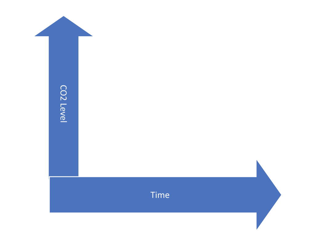
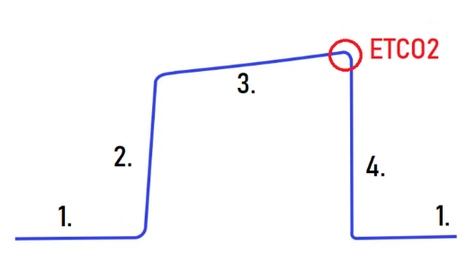
The capnography waveform is a simple graphical representation of quantitative CO2 levels on the Y axis and time on the X axis (See Figure 1). A normal capnography waveform is in a square shape with rounded corners (see Figure 2).
Figure 1: Capnography X- and Y- Axis
The Four Phases of Capnography
To best understand the graphical reading, it is important to understand what each phase of the capnogram represents.
Figure 2: Normal Waveform and Phases
Source: https://www.christienursing.com/post/etco2
- Phase 1: This phase is the beginning of exhalation, also known as the baseline, because there is no CO2 present at this point. In this phase, no gas exchange is occurring.
- Phase 2: CO2 from the lungs reaches the upper airway, which causes a rapid increase in CO2. This is called the ascending phase, or early exhalation.
- Phase 3: In this phase, CO2 is recorded consistently in the oral cavity, nose, and lungs.
- Phase 4/Phase 0: This is the end-tidal phase, which is also the start of inspiration. Graphically, this phase is the descending phase because inhalation begins, oxygen fills, and CO2 concentration decreases.
The PQRST of Capnography
To confirm placement of artificial airways and to monitor ventilation through waveform capnography, the clinician must be able to read and understand the PQRST – an acronym for Proper, Quantity, Rate, Shape, and Trend.
- Proper: This term simply means to know the normal readings for quantity, rate, shape, and trending of waveform capnography. These values are the same for healthy individuals regardless of age or sex.
- Quantity: Know the normal, quantifiable CO2 range (35-45 mmHg).
- Rate: The proper respiratory rate for adults is 12-20 breaths per minute (bpm). For ventilated adults, the rate should be slower, at about 10-12 bpm. Too rapid ventilation does not let enough CO2 build up in the alveoli, which results in lower end-tidal CO2 readings. Ventilating too slowly results in extra CO2 building up and an artificially high reading.
- Shape: Effective use of waveform capnography means knowing at a glance what the normal waveform should look like. Typical waveforms are rectangular with rounded corners. Various abnormal waveforms can be indicators of different health conditions.
- Trend: To determine effectiveness of treatments, and assess the patient’s condition, it is important to be able to trend the quantity, rate, and shape of waveform capnography readings. Ideally, these metrics should all be stable or improving.
The Five Characteristics of a CO2 Waveform
Understanding and interpreting capnography waveforms requires practice and a good understanding of the five waveform characteristics: height, frequency, rhythm, baseline, and shape. This information, once mastered, can be used for diagnosis and ventilator troubleshooting in ACLS and other emergency and critical care scenarios.
The five characteristics of a waveform are descriptors that are necessary to communicate changes. The following are examples of how changes in these characteristics are applied:
Hypoventilation
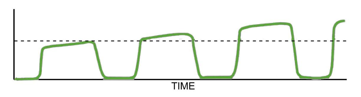
This condition occurs because of either slow or inadequate respirations. It leads to a buildup of carbon dioxide in the body, since not enough is expired with each breath. The waveform height will appear taller because the ETCO2 is above 45 mmHg. The waveform shape will change in that the rectangles will be stretched out due to the longer time between breaths.
Figure 3: Hypoventilation Waveform
Source: https://www.capnomask.com/what-is-waveform-capnography
Hyperventilation
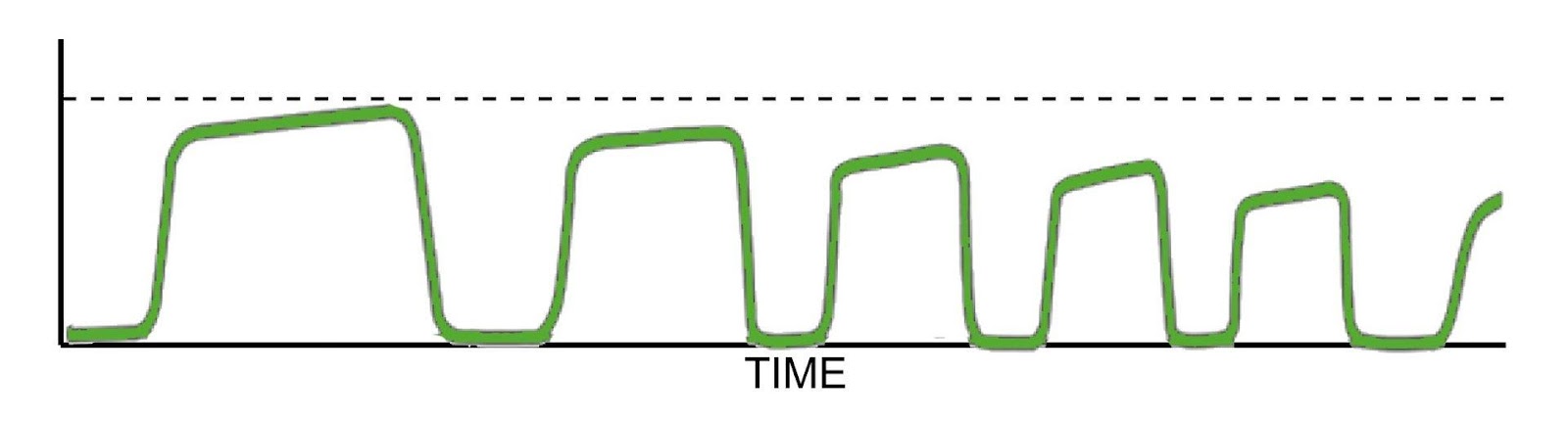
A rapid respiratory rate causes excess CO2 to be eliminated. This results in a decreased level of CO2 (below 35 mm Hg or below baseline measurements). There is also a shorter time between breaths and a higher frequency of breaths. The waveform will thus have smaller, shorter waveforms.
Figure 4: Hyperventilation Waveform
Source: https://www.capnomask.com/what-is-waveform-capnography
Obstructive Respiratory Conditions
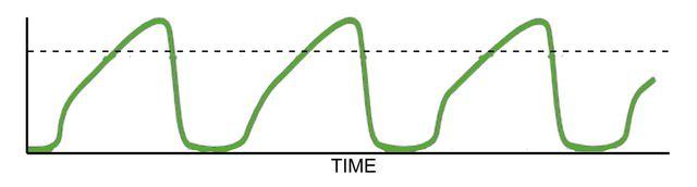
In the case of patients with diseases like asthma, emphysema, and COPD, the waveforms can look different. The phase three (top of the square) results can be more slanted, causing a “shark fin” shape. This shape occurs because of a struggle to expel CO2. A tall waveform can also result because CO2 is trapped, causing the level to be above 45 mmHg.
Figure 5: Obstructive Disease Waveform
Source: https://www.capnomask.com/what-is-waveform-capnography
There are many more patterns that clinicians learn to watch for as indicators of certain conditions. However, these conditions are the most commonly seen.
ACLS Applications for Waveform Capnography
Advanced capnography training will allow clinicians to recognize patterns that are associated with common issues. Below are some examples that most relate to ACLS:
Cardiopulmonary Resuscitation
Waveform capnography is very useful in monitoring the effectiveness of resuscitative efforts in the intubated patient.
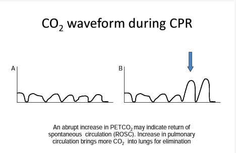
The waveform during CPR appears in a sawtooth pattern that is displayed at the rate of compressions. During CPR, the reading of concern via capnography is patient end-tidal CO2 (PETCO2). This reading is the maximum partial pressure of CO2 at the end of a breath. The effectiveness of compressions for CPR can be monitored graphically by ensuring that PETCO2 values remain between 10 and 20 mmHg. PETCO2 values less than 10 mmHg during ACLS are associated with poor patient outcomes.
While monitoring waveform capnography during resuscitation, an abrupt increase in PETCO2 may mean the return of spontaneous circulation (ROSC).
Figure 6: Waveform Capnography Monitoring during CPR
Source: https://www.capnography.com/images/pppdf/cprpdf.pdf
Endotracheal Tube Placement
Waveform capnography is known as the most reliable tool (outside of radiography) to confirm placement of advanced airways. When the endotracheal tube (ET Tube) is placed appropriately, the clinician will see an end-tidal CO2 reading appear almost immediately after ventilation starts.
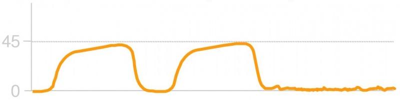
It is important to note that all capnography readings rely on air movement and circulation of CO2 to the lungs to produce a reading. Even in the case of correct ET tube placement, a waveform may not be produced if compressions are not fast or deep enough.
Figure 7: ET Tube Displaced
Source: https://www.roaddoc.com/scems/index.php/End_Tidal_CO2_Monitoring_and_Capnography
Studies have shown that capnography is superior to checking for bilateral breath sounds or the visualization of cords to confirm placement of ET tubes in emergency situations. Because of this research, the American Heart Association recommends the following to medical professionals:
- Monitor capnography and breath sounds in the initial stages of resuscitation to establish the baseline.
- Compare these early readings to subsequent readings after airway placement.
- Confirm correct ET tube placement by observing for capnography waveforms, listening to confirm the absence of epigastric sounds, and an ETCO2 level the same or higher than baseline.
- At any time during resuscitation, the loss of ETCO2 after the first ventilation indicates that the airway is probably misplaced or dislodged.
- An ETCO2 reading and waveform that suddenly decreases may indicate that the ET tube has been placed too deep and has migrated to the right bronchus. This should be assessed by listening for the absence of air movement over the left lung.
Troubleshooting Tips
Troubleshooting tips for waveform capnography are closely aligned with the methods used to troubleshoot airways. A simple mnemonic called DOPE is often used to guide the assessment – dislodgement, obstruction, pneumothorax, and equipment failure.
When ETCO2 is suddenly lost in an intubated patient, the first action is to check a pulse. Just like an immediate rise in ETCO2 can indicate ROSC, the loss of circulation will cause an immediate drop. If this cause is ruled out, then proceed to use DOPE.
- Assess the patient’s bilateral breath sounds to check for a dislodged tube, such as during a right bronchus placement.
- Suction the tube to assess for obstruction, such as with a mucous plug.
- Also listen to detect a possible pneumothorax (absent breath sounds on the affected side, indicating possible tracheal deviation).
- If all clinical signs have been ruled out, then assess for equipment problems with the capnography circuit itself such as pinched tubing, detached lines, or a malfunctioning machine.
Conclusion
With a good understanding of waveform capnography, ACLS-trained clinicians will be even more prepared to provide high-quality resuscitation and monitor progress. Capnography is a valuable tool to provide high-quality care, whether it is used in a hospital or EMS setting.
If you would like to learn more about the drugs, ECG rhythms, clinical scenarios, and other topics related to ACLS, AMRI has study materials that can help you deepen your understanding. Accredited by the National Board of Emergency Care Certifications (NBECC), AMRI has helped more than one million medical professionals earn their ACLS, BLS, and PALS certifications or recertifications since 1983.
Register for your exam with AMRI today to gain access to premium study materials and our widely accepted certification program.